


You've found a lump in your breast, pressed it again to make sure it's really there, and now your mind is racing. That reaction is completely understandable. What you need in that moment isn't more alarming headlines. You need clear, calm guidance on what a moving lump can mean and what to do next.
The first reassuring point is that most breast lumps are not cancer. High-quality clinical sources estimate that roughly 80% of biopsied breast lumps are benign, and for women in their 30s, the chance that a palpable breast lump is cancer has been estimated at about 1 in 204 (0.49%) according to Stony Brook Medicine's breast lump overview. That matters because fear often fills in the gaps before facts do.
What helps most is shifting away from self-diagnosis and towards a proper assessment. If anxiety is spiralling while you wait to be seen, practical strategies for coping with overthinking and anxiety can help you stay grounded without ignoring the symptom.
Finding a Breast Lump and Understanding What Comes Next
A lump that moves under your fingers often turns out to be benign. Even so, touch alone isn't enough to tell what it is. A lump can feel mobile and still need proper imaging, and that's exactly why breast clinics rely on a structured process rather than guesswork.
Why panic doesn't help, but action does
When patients first notice lumps in breast that move, they often ask the same question: “If it moves, does that mean it's harmless?” The honest answer is that mobility is reassuring, but not definitive. It points more often towards common benign causes, especially in younger women, but it doesn't replace an examination.
A more useful question is: what's the right next step? In UK practice, that usually means arranging a GP review rather than watching and waiting in uncertainty.
Practical rule: A breast lump deserves an assessment because the goal isn't to guess correctly at home. The goal is to reach a diagnosis safely and promptly.
What your doctor is trying to work out
A clinician doesn't look at one feature in isolation. They build a picture from several details, including:
- How long it's been there. A brand-new lump is treated differently from a long-standing unchanged one.
- Whether it changes. Growth, increasing firmness, or a change in surrounding tissue matters.
- What else is happening. Pain, nipple change, skin dimpling, redness, or thickening all add context.
- Your age and breast density. These affect which scan is most useful.
That process often reassures patients quickly, because uncertainty is usually the hardest part. Once you've booked an appointment, you've already moved from fear into action.
What not to do while waiting
Several habits make the waiting period worse:
- Repeatedly checking the lump. This can make the area sore and doesn't improve accuracy.
- Searching for one “perfect” symptom match online. Breast lumps don't read textbook descriptions.
- Assuming pain means benign or no pain means cancer. Neither rule is reliable.
- Delaying because it seems to move. Mobility is one piece of information, not a diagnosis.
The most sensible approach is simple. Notice the change, book the appointment, and make a few notes so you can describe it clearly.
What Are Most Movable Breast Lumps?
A mobile lump is technically most consistent with a benign lesion such as a fibroadenoma or a cyst, both of which are classically smooth, round, and easily moved under the skin. Fibroadenomas are the commonest benign solid breast tumour in younger women, and UK breast-imaging pathways typically use ultrasound first for a palpable mass in younger patients to distinguish solid from fluid-filled lesions, as outlined in the Merck Manual overview of breast lumps.
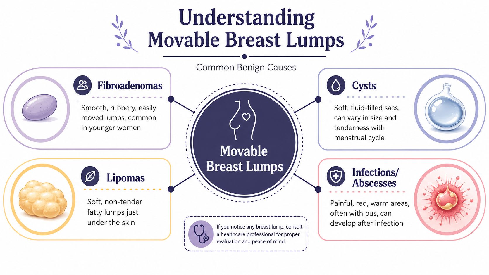
Fibroadenomas
A fibroadenoma often feels smooth, rubbery, and easy to nudge under the skin. Some patients describe it as a small object that slips away from their fingers. That mobile quality is one reason it causes so much confusion. It feels distinct, but not fixed.
These lumps are especially common in younger women. In practice, when a younger patient has a smooth, mobile breast lump, fibroadenoma is often high on the list of possibilities. That doesn't mean it should be assumed. It means ultrasound is particularly useful for clarifying what's there.
Cysts
A cyst is a fluid-filled sac. It can feel softer than a fibroadenoma, although deeper cysts can sometimes feel firmer than expected. Some become more noticeable or more tender around the menstrual cycle, which can make them seem to appear suddenly.
What works here is observation with purpose, not guesswork. If a lump seems to fluctuate with your cycle, note that down and mention it. That detail can help a clinician interpret the finding, but it still shouldn't replace proper review.
A moving lump often has a benign explanation, but the useful question is not “What do I hope it is?” It's “What test will tell us for sure?”
Other possible benign causes
Not every mobile lump is a fibroadenoma or cyst. Other benign possibilities include:
- Lipoma. A soft fatty lump just under the skin that can feel squashy and mobile.
- Infection or abscess. More likely if the area is red, warm, painful, or associated with feeling unwell.
- Hormonal lumpiness. Some breasts feel generally nodular rather than having one distinct mass.
These possibilities behave differently. A lipoma is usually soft and unobtrusive. An infected area is more likely to be tender and inflamed. Hormonal lumpiness often feels broader and less defined than one discrete lump.
What helps patients most
What works is describing the lump as clearly as you can:
| Feature | What to notice |
|---|---|
| Movement | Does it slip under your fingers or feel anchored? |
| Texture | Smooth, rubbery, soft, or hard? |
| Shape | Round, oval, or irregular? |
| Timing | Has it changed around your period? |
| Symptoms nearby | Any redness, warmth, skin change, or nipple change? |
That sort of description is far more useful than trying to name the lump yourself.
Benign Versus Concerning Signs to Be Aware Of
In UK clinical practice, a new breast lump that is mobile is still investigated as potentially malignant because palpability alone is not discriminatory. The NHS advises urgent assessment for a lump or thickening in the breast, and imaging or diagnostic work-up is required rather than relying on touch, as summarised in the Cleveland Clinic breast lump overview reflecting NHS-style assessment principles.
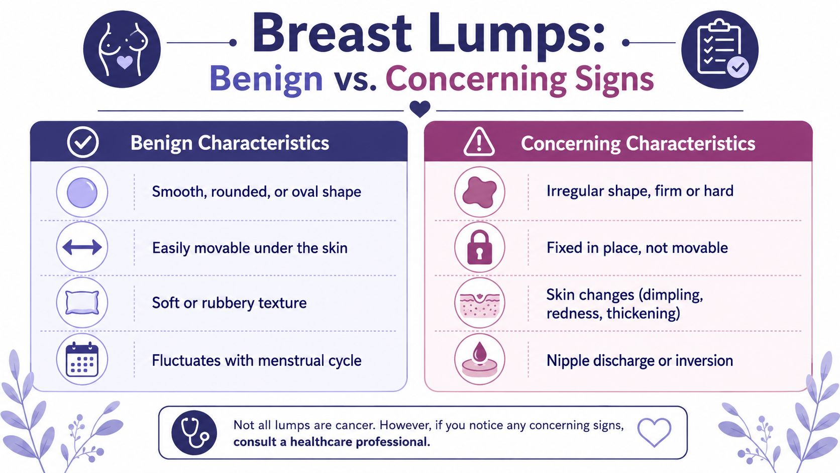
Features that often look more reassuring
Some lump features lean more towards a benign explanation. They don't prove it, but they're useful clues.
- Smooth outline. Benign lumps are often rounded or oval rather than irregular.
- Easy mobility. A lump that moves freely under the skin is often less concerning than one that feels tethered.
- Soft or rubbery feel. Many harmless lumps aren't rock hard.
- Cycle-related change. If it varies with menstruation, a hormonal or cystic cause becomes more plausible.
Features that need faster attention
Other changes should lower your threshold for seeking prompt review.
- Fixed position. A lump that feels stuck to surrounding tissue deserves careful assessment.
- Firm or irregular feel. Hard, uneven lumps raise more concern.
- Skin change. Dimpling, puckering, thickening, redness, or a change in breast contour matters.
- Nipple change. New inversion or discharge should be mentioned.
- Progressive change. If it's enlarging or becoming more obvious, don't sit on it.
The trade-off patients often miss
People often want a binary answer. Smooth and moveable means fine. Hard and fixed means bad. Real life isn't that tidy.
Some benign lumps feel worrying. Some concerning lumps don't fit the classic picture. That's why a checklist can guide your thinking but can't replace the clinic pathway.
Key point: No single sign, including mobility, can rule cancer in or out.
A practical comparison
| More often benign | More concerning |
|---|---|
| Smooth and well-defined | Irregular or poorly defined |
| Mobile under the skin | Fixed in place |
| Soft or rubbery | Firm or hard |
| Fluctuates with cycle | Progressively enlarging |
| No skin or nipple change | Skin dimpling or nipple change |
Use this as a prompt for what to report, not as a home test to rely on.
What patients should bring to the appointment
A short note on your phone is enough. Include:
- When you first noticed it
- Whether it has changed
- Whether it’s painful
- Any relation to your menstrual cycle
- Any skin or nipple changes
That usually leads to a better consultation than trying to remember everything in the room.
The Path to Clarity What to Expect During an Evaluation
The most helpful part of a breast lump assessment is often the structure. Once you know the usual pathway, the experience feels less mysterious and much more manageable.
For younger patients and for women with denser breast tissue, imaging choices matter. A UK clinical review notes that in women under 35, ultrasound is typically preferred, while after 35 mammography becomes the investigation of choice and is less sensitive in dense breasts, which is why specialist assessment is so important, as explained in this UK review of breast disease assessment.
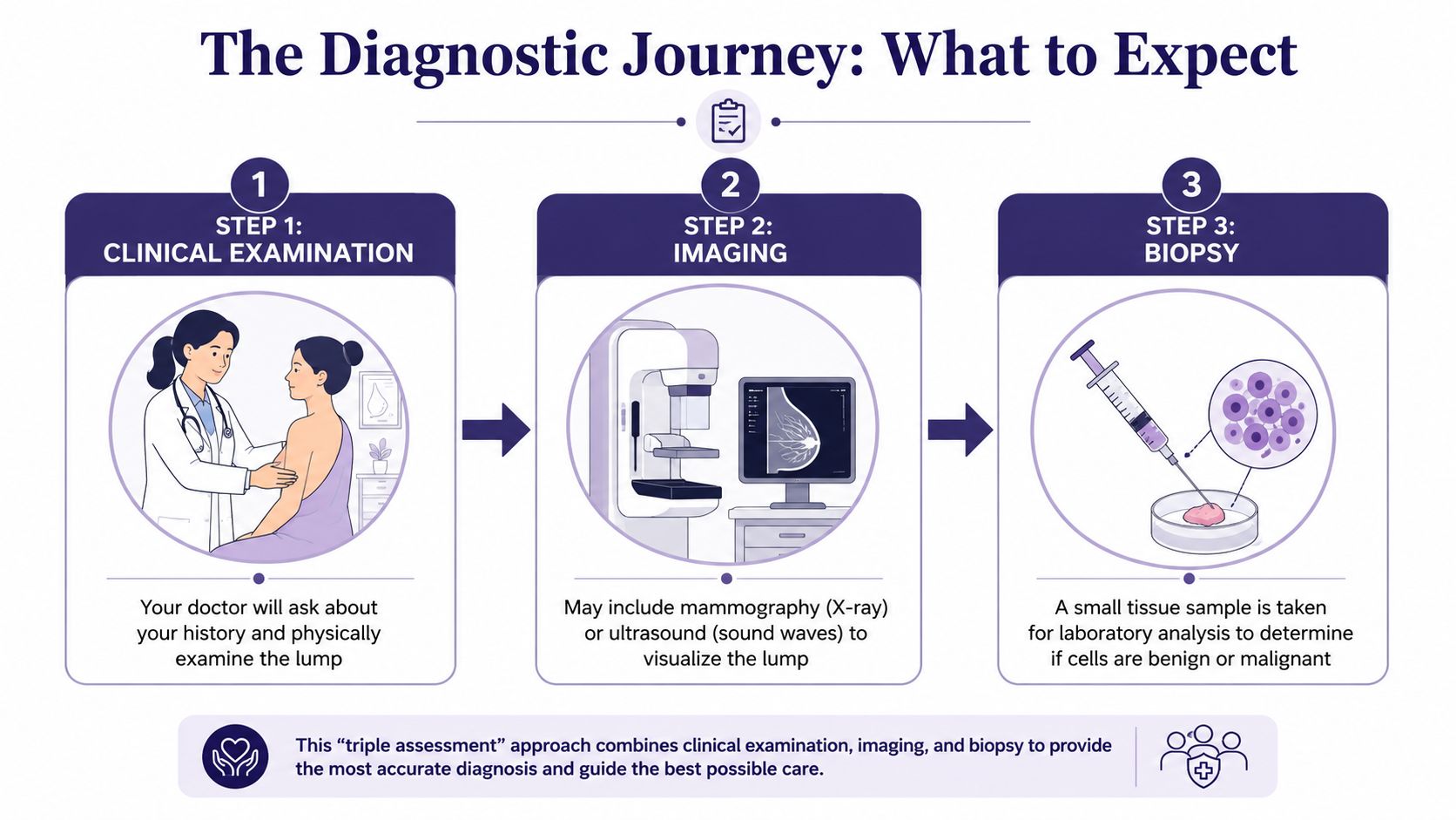
Step one is the clinical examination
At the first appointment, your doctor usually asks focused questions. They’ll want to know when the lump appeared, whether it changes with your cycle, whether there’s pain, and whether you’ve noticed any nipple or skin changes.
The examination itself is practical and systematic. A clinician assesses the lump’s size, shape, mobility, consistency, and whether there are associated changes in the surrounding breast or underarm area.
Step two is imaging
Imaging gives the first real clarification. In younger patients, ultrasound is often the most useful starting point because it can distinguish a fluid-filled cyst from a solid lump. In older patients, mammography is more commonly used, though dense breast tissue can make interpretation trickier.
At this point, self-diagnosis usually falls away. A lump that felt “obviously movable” at home may turn out to be a cyst, a fibroadenoma, or something that needs further sampling.
If you’re ever called back after breast imaging, it can help to understand that a callback doesn’t automatically mean cancer. A calm explanation of the process in navigating a callback mammogram notice can reduce a lot of unnecessary panic.
Step three is biopsy if needed
Not everyone needs a biopsy. If imaging looks clearly benign, your doctor may recommend reassurance or follow-up instead. If the picture remains uncertain, a biopsy takes a small sample of tissue so the cells can be examined properly.
That’s the key trade-off. Imaging is excellent, but when the answer still isn’t clear, a tissue sample provides certainty that touch and scans alone sometimes can’t.
The point of triple assessment is simple. Each step answers a different question, and together they reduce the chance of false reassurance.
Private assessment and faster decision-making
One advantage of private care is speed. If delays are adding to the worry, some patients choose to explore private cancer screening options so they can move more quickly from “I’ve found something” to “I know what it is”.
That matters because uncertainty tends to grow in the gaps between appointments. Clear timelines reduce stress almost as much as good explanations do.
Proactive Lifestyle Habits for Your Breast Health
Lifestyle won’t diagnose a breast lump, and it won’t make a suspicious one safe to ignore. What it can do is support your overall health, improve breast awareness, and make it easier to spot genuine change early.

Focus on breast awareness, not obsessive checking
The most useful habit is knowing what’s normal for your own body. Breasts can feel naturally lumpy, and that texture can shift across the month. Patients who know their usual pattern tend to notice meaningful changes sooner.
What doesn’t work is checking repeatedly because you’re frightened. Frequent poking makes tissue sore and can make a normal area feel more prominent.
A simple self-check routine
Choose a consistent time each month. If you have periods, many people find it easiest to check after the cycle has settled.
Try this approach:
- Look first. Stand in front of a mirror with arms relaxed, then raised. Notice any visible contour change, puckering, or nipple change.
- Feel in the shower or lying down. Use the flat pads of your fingers, not the fingertips.
- Cover the whole breast. Move in a pattern you can repeat each month, such as small circles from the outer breast towards the nipple.
- Include the underarm area. Breast tissue extends beyond the breast itself.
- Note what’s different. One distinct new lump is more important than general lumpiness you always have.
Day-to-day habits that support health
There’s no single “breast health diet”, but a few broad habits are sensible and sustainable:
- Stay active. Regular movement supports weight management, energy, and metabolic health.
- Aim for a balanced diet. Build meals around vegetables, fruit, fibre, protein, and minimally processed foods.
- Limit alcohol. If you drink, keeping intake modest is a sensible health decision.
- Prioritise sleep and stress management. Poor sleep and chronic stress don’t cause a lump directly, but they make symptoms feel harder to manage and can disrupt healthy routines.
- Seek support for hormone-related symptoms. If breast tenderness or lumpiness seems strongly cycle-linked, broader guidance on natural hormone balance for women may be a helpful starting point for lifestyle questions alongside medical advice.
What actually helps in practice
A short monthly habit beats an intense burst of checking after a scare. It is often more effective to use a recurring reminder on a phone and a simple note of any change.
Know your baseline. Then act on change.
That combination is much more useful than trying to perform a perfect self-exam.
When to See a Doctor and How to Get Rapid Answers
The key clinical point is straightforward. Mobility alone does not rule out cancer. A lump that is new, enlarging, or present for more than 4 to 6 weeks is usually referred for assessment rather than merely observed, as outlined in the Mayo Clinic guidance on suspicious breast lumps.
Book an appointment if any of these apply
You shouldn’t wait for a lump to become dramatic before getting help. Arrange a review if you notice:
- A new lump that you haven’t felt before
- A lump that’s growing or becoming more obvious
- A persistent lump that hasn’t settled over several weeks
- Skin changes such as dimpling, thickening, or redness
- Nipple change including inversion or discharge
- A fixed or hard area that feels different from the surrounding tissue
If you’re pregnant, breastfeeding, postmenopausal, or unsure what you’re feeling, the answer is the same. Get it checked.
What rapid access changes
The hardest part for many patients isn’t the test itself. It’s the waiting. Long gaps between finding a symptom, seeing a doctor, and getting imaging can make even a benign situation feel unmanageable.
Faster assessment gives you two things. First, it reduces the chance of delay if something needs treatment. Second, it shortens the period where your imagination is doing the work that imaging should be doing.
For patients trying to find a timely appointment locally, seeing a private GP can be a practical way to get examined promptly and referred on for imaging without the usual backlog.
What to do today
If you’ve found lumps in breast that move, don’t try to solve the puzzle alone. Use this simple plan:
- Write down the change
- Stop repeated checking
- Book a GP appointment
- Attend imaging if advised
- Follow through until you have a clear answer
That’s the part many articles miss. Reassurance doesn’t come from guessing that a lump is probably benign. It comes from completing the assessment properly.
If you want prompt, thoughtful assessment without feeling rushed, The Lagom Clinic offers private GP appointments in Bristol with time to discuss your symptoms properly, examine the concern carefully, and arrange rapid onward investigation when needed. When you’ve found a breast lump, getting clear answers quickly can make all the difference.


 Members Consultation