


When your doctor recommends an imaging scan, you might hear the terms ‘CT’ and ‘MRI’ used. While both give a detailed look inside your body, they are fundamentally different tools, each with its own strengths.
Think of it like this: a CT scan uses X-rays to create incredibly quick, detailed pictures of bones, organs, and blood vessels. This makes it a workhorse in emergency situations. In contrast, an MRI uses powerful magnets and radio waves to generate stunningly clear images of soft tissues—things like muscles, ligaments, and the brain. It's the go-to for diagnosing complex conditions where fine detail is paramount.
Understanding Your Imaging Options: CT Scan vs. MRI
Knowing the difference between a CT (Computed Tomography) scan and an MRI (Magnetic Resonance Imaging) scan helps you take a more active role in your healthcare. They might sound similar, but they work in completely different ways and are selected for very specific clinical reasons.
A CT scan—sometimes called a CAT scan—is essentially a sophisticated, 3D X-ray. It’s brilliant at showing dense structures like bone with exceptional clarity. Its speed is its superpower, making it invaluable in urgent scenarios like pinpointing internal bleeding after an accident, diagnosing a stroke, or evaluating a complex fracture.
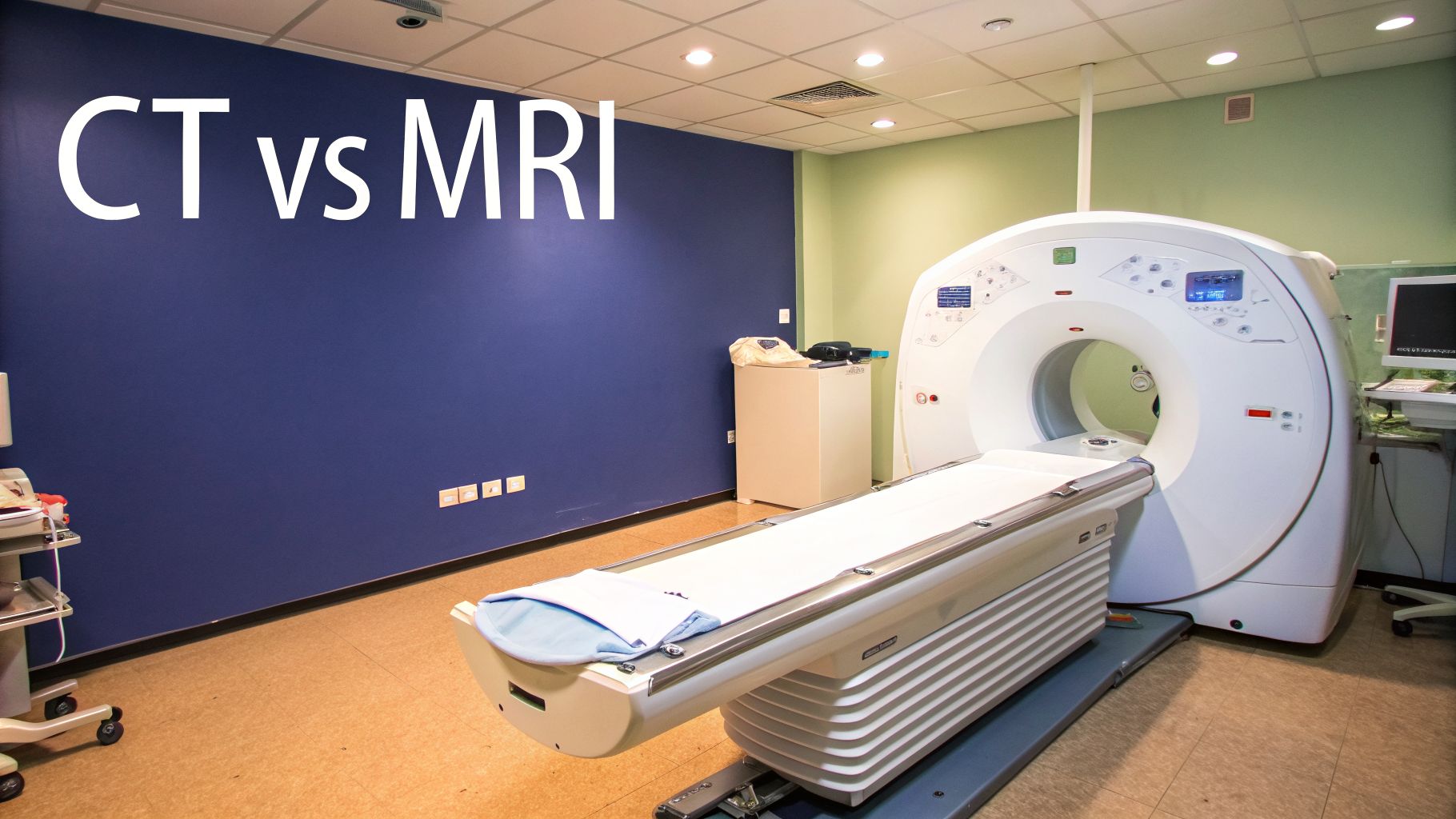
An MRI, on the other hand, gives an unmatched view of the body’s soft tissues. It can pick up on subtle details a CT might miss, like a tiny tear in a knee ligament, a herniated disc pressing on a nerve, or the earliest signs of a brain tumour. The price for this incredible detail is time; an MRI scan takes quite a bit longer to complete.
CT Scan vs MRI: Key Differences at a Glance
To help you and your doctor decide on the best path forward, it’s useful to see a direct comparison. This table breaks down the essential distinctions between these two powerful diagnostic tools.
| Feature | CT (Computed Tomography) Scan | MRI (Magnetic Resonance Imaging) Scan |
|---|---|---|
| Technology | Uses specialised X-rays to create cross-sectional images. | Uses powerful magnets and radio waves to build a detailed picture. |
| Best For | Bone fractures, internal injuries, lung and chest issues, cancer detection. | Soft tissue injuries (ligaments, tendons), brain and spinal cord issues, joint problems. |
| Scan Time | Very fast, typically under 15 minutes. | Slower, often taking 30 to 60 minutes or more. |
| Radiation | Involves a small, controlled dose of ionising radiation. | No ionising radiation is used. |
| Patient Experience | Open, doughnut-shaped machine; generally less likely to cause claustrophobia. | Enclosed tube-like machine, which can be challenging for some patients. |
Ultimately, the choice isn't about which scan is "better," but which one is right for the specific medical question at hand.
A CT scan is the clear winner for a rapid trauma assessment in A&E, whereas an MRI is superior for getting a meticulous look at what’s going on inside your knee joint.
Having this foundational knowledge makes it easier to understand your doctor’s recommendation. The goal is always the same: to get the clearest possible picture of your health concern in the safest, most effective way. By appreciating their unique strengths, you can feel much more confident about the diagnostic journey ahead.
How Each Scan Actually Works
On the surface, CT and MRI scans seem similar—they both give doctors a detailed look inside your body. But how they get that picture is what really sets them apart. Think of it less like choosing between two brands of the same thing and more like choosing between a high-tech camera and a sophisticated mapping tool.
One uses X-rays to rapidly build a 3D picture, almost like a digital sculptor carving out an image of your insides. The other uses a massive magnet to gently coax signals from the water in your body's cells. Knowing the difference helps you understand why your doctor might pick one over the other and makes the whole process feel a bit less mysterious.
The Mechanics of a CT Scan
A Computed Tomography (CT) scan—or CAT scan, as you might have heard it called—is basically an X-ray on steroids. A standard X-ray gives you a single, flat image, a bit like a shadow. A CT scanner, however, takes this a huge step further.
The machine’s X-ray source spins around you, taking hundreds, sometimes thousands, of pictures from different angles. It’s a bit like taking photos of an object from every conceivable viewpoint. A computer then stitches all these individual ‘slices’ together to create an incredibly detailed 3D model.
This method is brilliant for showing differences in the density of tissues, which is why it’s the go-to for:
- Bone: It can show up tiny, complex fractures or wear-and-tear that a normal X-ray would miss. Maintaining good bone health through calcium-rich foods and weight-bearing exercise can reduce fracture risk.
- Lungs: Perfect for spotting nodules, infections, or dangerous blood clots. Lifestyle choices like quitting smoking and regular cardiovascular exercise are crucial for lung health.
- Blood Vessels: With a bit of contrast dye, it can light up blockages or aneurysms. A heart-healthy diet low in saturated fats can help keep blood vessels clear.
Its biggest advantage is speed. A scan can be over in minutes, which is why CT is a cornerstone of A&E departments for assessing major trauma, suspected strokes, or internal bleeding where every second counts.
A good way to picture a CT scan is to imagine a loaf of sliced bread. A regular X-ray shows you the whole loaf from the outside. A CT lets you pull out each slice and inspect it individually, seeing everything hidden inside.
The Science Behind an MRI Scan
Magnetic Resonance Imaging (MRI) works in a completely different way and doesn't use any ionising radiation at all. Instead, it relies on a very strong magnetic field and radio waves. It all comes down to the water in your body.
Your body is about 70% water, and every water molecule has hydrogen protons. When you slide into the MRI machine, the powerful magnet makes all these tiny protons line up in the same direction, like microscopic soldiers on parade.
Next, the machine sends a quick pulse of radio waves into the specific area being scanned, knocking the protons out of alignment. As soon as the pulse stops, the protons snap back into line, releasing a tiny energy signal as they do. Different types of tissue—like muscle, fat, and nerve—have different amounts of water, so their protons realign at different speeds.
A sophisticated receiver picks up these signals, and a computer uses them to create a stunningly detailed picture. This makes MRI the undisputed champion for visualising soft tissues, giving unparalleled clarity on:
- Muscles, ligaments, and tendons
- The brain and spinal cord
- Internal organs like the liver or kidneys
Why a Contrast Agent Might Be Used
Sometimes, to get an even clearer picture, the radiologist will use a special dye called a contrast agent. This isn’t a dye in the traditional sense; it’s a substance that temporarily makes certain tissues stand out on the scan, helping to highlight exactly what’s going on.
For a CT scan, this is usually an iodine-based contrast. It’s injected into a vein and circulates through your bloodstream, making your blood vessels and organs with a rich blood supply (like your liver) appear bright white on the final image. This is a game-changer for spotting tumours, inflammation, or problems with blood flow.
For an MRI, the approach is different. Radiologists use a gadolinium-based contrast agent. Gadolinium works by interacting with the water molecules at a magnetic level, making them stand out more. It’s incredibly useful for getting a better look at tumours, checking for inflammation in joints, or getting the clearest possible view of the brain and spinal cord. Your doctor will always run through the pros and cons of using contrast before you have your scan.
When Your Doctor Might Recommend a CT Scan or an MRI
It can feel a bit confusing when your doctor recommends a scan, but understanding why they’ve chosen a CT or an MRI can make all the difference. The decision isn’t random; it boils down to two key things: how quickly we need answers, and whether we’re looking at hard structures like bone or soft tissues like ligaments and organs.
By walking through a few real-world examples—from a sudden accident to a nagging sports injury—you’ll see the logic behind each choice. It helps put the procedure into context and shows how it fits into your broader health picture.
Scenarios Where a CT Scan Is the Best Choice
A CT scanner’s greatest strengths are its incredible speed and its ability to create detailed images of bone, organs, and blood vessels. This makes it the go-to tool in urgent or even life-threatening situations where doctors need clear answers, fast.
Here are a few classic examples:
- Assessing Head Trauma: After a car crash or a nasty fall, a CT scan of the head is the priority. It takes just a few minutes and is the quickest way to spot a skull fracture, brain swelling, or internal bleeding—all of which need immediate attention.
- Diagnosing Acute Abdominal Pain: If you show up at A&E with sudden, severe stomach pain, a CT scan can swiftly get to the bottom of it. It can reveal conditions like appendicitis, kidney stones, or a bowel obstruction, giving surgeons a clear roadmap for what to do next. To support good digestive health, focus on a high-fibre diet and stay well-hydrated.
- Staging Cancer: When someone is diagnosed with cancer, a CT scan of the chest, abdomen, and pelvis helps doctors see if it has spread to other parts of the body. This rapid ‘staging’ process is crucial for putting together the most effective treatment plan.
The numbers back this up. In England, the use of CT scans has shot up by a massive 60% between 2016/17 and 2023/24, growing much faster than MRI usage. This really highlights its vital role in urgent care, especially when time is of the essence. You can read more about these diagnostic imaging trends and what they mean for patient care.
When an MRI Is the Right Tool for the Job
While CT scans are the heroes of the emergency room, an MRI is the specialist for detailed, non-urgent investigations of soft tissues. Its real magic is producing incredibly high-resolution images of muscles, nerves, and joints without any radiation, making it perfect for diagnosing chronic conditions and subtle injuries.
Think about these situations where an MRI provides the clarity needed:
- Diagnosing a Runner's Knee Injury: If you’re a runner with knee pain that just won’t quit, an MRI gives a crystal-clear picture of your ligaments, tendons, and cartilage. It can spot a tiny tear in the meniscus or ACL that a CT scan would miss. Strengthening supporting muscles and proper stretching can help prevent such injuries.
- Investigating Back Pain and Spinal Issues: For long-term back pain or sciatica, an MRI offers an unmatched view of the spinal cord, nerves, and the discs between your vertebrae. It can clearly show a herniated disc pressing on a nerve. Maintaining a strong core through exercises like Pilates can provide crucial support for your spine.
- Getting a Clear View of Brain Health: When symptoms are less urgent but still worrying—like persistent headaches, dizziness, or small memory changes—a brain MRI is the gold standard. It can detect conditions like multiple sclerosis, small tumours, or the effects of a minor stroke. A diet rich in omega-3s and regular exercise are known to support long-term brain health.
This simple flowchart helps to visualise when each scan is typically chosen.
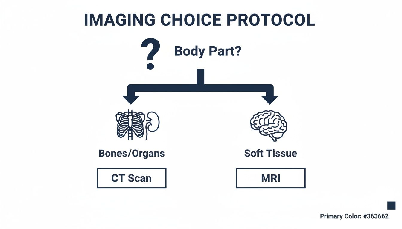
As the chart shows, it's a fundamental principle: CTs are generally the first pick for looking at bones and solid organs, while MRIs are the best choice for diving deep into soft tissues.
The choice between a CAT scan vs MRI is never arbitrary. It’s a calculated decision based on your specific symptoms, the body part involved, and the level of detail required to get you the right diagnosis and treatment.
Knowing the difference between these powerful tools helps you understand why your GP might recommend one over the other. It puts you in a much better position to have an informed conversation about the next steps in your care.
Getting Ready for Your Scan: Safety Checks and Practical Tips
Knowing what to expect before you go in for a CT or MRI scan can make a world of difference. It helps calm the nerves and ensures everything goes off without a hitch. A bit of preparation, from safety checks to small lifestyle tweaks, puts you in control and helps us get the clearest possible images while keeping you safe.
The first thing to understand is the core safety difference between the two. With a CT scan, the main consideration is radiation. For an MRI, it's all about that powerful magnetic field. Getting your head around these two points helps demystify the whole process.

CT Scans and Radiation: A Balanced View
It’s completely normal to have questions about the radiation involved in a CT scan. The good news is that modern scanners are incredibly efficient, using low-dose techniques to get high-quality images with the absolute minimum radiation required.
When it comes down to it, the diagnostic benefit of pinpointing the cause of your symptoms almost always outweighs the very small risk. For perspective, the dose you receive is often comparable to a few years of the natural background radiation we’re all exposed to just by living on planet Earth. Your clinical team will always weigh this up and only recommend a scan when it’s truly necessary for your diagnosis.
Why MRI Safety Screening is So Important
An MRI scanner houses an incredibly powerful magnet, which means the safety screening process is absolutely critical. We don’t take any shortcuts here. This detailed check is designed to protect you from any harm that could be caused by the magnetic field interacting with metal on or inside your body.
Before your scan, you’ll need to fill out a comprehensive safety questionnaire. It is vital that you answer everything honestly and completely. You must tell the radiographer if you have any of the following:
- A pacemaker or implantable defibrillator (ICD)
- A cochlear implant
- Certain types of clips used for brain aneurysms
- Metal coils placed within your blood vessels
- Any history of metal fragments in your body, especially near your eyes
The powerful magnet in an MRI scanner is always on. That's why the safety checks are so rigorous—it’s not just for patients but also to protect staff from the danger of metallic objects being pulled toward the machine.
Simple Steps to Prepare for Scan Day
Beyond the specific safety rules for a CAT scan vs MRI, a few practical tips can make your experience much smoother. These are simple things to prepare you both physically and mentally for the appointment.
Hydration is your best friend. Drinking plenty of water in the days before your scan is a great idea, particularly if you’re having a contrast agent. It makes your veins easier to find for the IV line and helps your kidneys flush the contrast out of your system afterwards.
You’ll also get specific instructions about food and drink. For certain scans, like those looking at the abdomen or when contrast is involved, you may be asked to fast for a few hours. Always stick to the guidance the clinic gives you.
Tackling Scan Anxiety and Claustrophobia
Feeling a bit anxious about a medical scan is incredibly common, especially with the enclosed nature of an MRI machine. The good news is there are proven techniques to help you stay calm and in control.
Deep breathing exercises are a surprisingly powerful tool. A great one to try is the 4-7-8 technique: breathe in quietly through your nose for 4 seconds, hold your breath for 7 seconds, and then exhale slowly through your mouth for 8 seconds. Practising this before you arrive can make it a go-to relaxation method during the scan.
Knowing exactly what to expect can also ease that feeling of uncertainty. For more details on the process and timing, have a read of our guide on how long an MRI takes.
Accessing Diagnostic Imaging in the UK
If you need a diagnostic scan in the UK, you have two main options: going through the NHS or opting for private healthcare. Both have their place, and figuring out which is right for you often comes down to balancing urgency against cost. Whether you're dealing with an injury or just want peace of mind, it helps to know how each system works.
The NHS offers fantastic care, but it's no secret that the system is stretched. Your journey typically starts with your GP, who will refer you for a scan if they deem it medically necessary. The catch? Waiting lists for non-urgent scans can be long, sometimes stretching into weeks or even months.
The Reality of UK Imaging Infrastructure
This strain isn't just about demand; it's also about resources. The UK has just 10 CT scanners per million people, which is well below the OECD EU average of 20.5 and fewer than any other EU country. The story is similar for MRI, with only 8.6 scanners per million people compared to the EU average of 12.4.
Add to that a projected 40% shortfall of clinical radiologists by 2028, and it’s clear why delays happen. You can read a full analysis of this from the BMA on how these pressures affect NHS diagnostics. For anyone with persistent pain or needing a diagnosis to plan a surgery, waiting simply isn't an option. This is where the private route becomes a real alternative.
Exploring the Private Imaging Route
Going private is a much more direct way to get the answers you need. At a clinic like The Lagom Clinic, the entire process is designed for speed and convenience, letting you bypass those NHS queues.
The main advantage is how quickly things move. You can often book a scan within a few days and get your results—along with a detailed report from a specialist radiologist—in as little as 24 to 48 hours. This kind of speed is crucial when you're worried about your health and need to make decisions about what to do next. To get a better idea of what proactive screening looks like, have a look at our guide on a full body scan.
Opting for private imaging is about more than just speed; it’s about taking control of your health journey. It allows you to schedule a scan at a time that suits you and get clear, actionable results without delay.
Costs and Insurance Considerations
Of course, the big question with private healthcare is always cost. The price of a scan can vary based on the type (cat scan v mri), the part of the body being imaged, and whether you need a contrast dye. As a general guide, a private MRI in the UK costs between £300 and £800, while a CT scan might range from £400 to £900.
Many people use private medical insurance to foot the bill. If this is your plan, it’s vital to check your policy details carefully. Spending a bit of time understanding policy exclusions beforehand can save you from any nasty surprises. By getting a clear picture of your options, you can choose the path that best fits your health needs, timeline, and budget.
Making an Informed Choice About Your Health
Trying to figure out medical imaging can feel a bit overwhelming, but when you grasp the fundamental differences between a CT scan and an MRI, you're in a much better position to be an active partner in your own healthcare. The key thing to remember is that one isn't simply 'better' than the other; it’s all about which one is the right tool for the job. That decision really comes down to a few critical factors.
The urgency of your situation, the specific part of the body being looked at, and what your doctor suspects might be going on will all point towards either a CT or an MRI. As we’ve covered, a CT scan is the go-to when time is of the essence—think trauma, suspected internal bleeding, or complex bone fractures. On the other hand, an MRI is what we turn to for incredible detail in soft tissues, making it the clear choice for joint injuries, spinal issues, and detailed brain assessments.
Taking a Proactive Role in Your Wellness
Knowing the difference between a cat scan v mri means you can have a much more productive chat with your GP. This partnership is the bedrock of a solid wellness plan that goes well beyond a single scan result. Your imaging report is a crucial piece of the puzzle, but it truly shines when it’s used to build a bigger picture of your health.
Think of your diagnostic results as a starting point for positive changes. Armed with the information from your scan, your doctor can give you specific, medically sound advice on:
- Nutrition: Adjusting your diet to support bone density, calm inflammation, or boost organ function.
- Physical Activity: Prescribing exercises to strengthen the muscles around an injured joint or to improve your heart health.
- Stress Management: Offering techniques to help you cope with chronic pain or lessen the physical toll stress takes on your body.
Your scan isn't just a picture; it's a launchpad for a conversation about your long-term health. It gives you the clarity to create a personalised plan that tackles root causes, not just the symptoms.
Building Your Comprehensive Health Plan
Even with significant investment, getting diagnostic tests done in a timely manner can still be a challenge. While NHS England's Community Diagnostic Centres (CDCs) have impressively delivered over 6.1 million tests since they started, the transition is slow. By August 2024, only 8% of CTs and 13.5% of MRIs were actually being performed in these new centres. This tells us there's still a heavy reliance on busy hospitals, which can mean delays. You can read more about the progress of community diagnostic centres.
This is often where choosing private access can give you peace of mind. By taking control of your diagnostic journey, you get the answers you need without the lengthy wait, letting you and your doctor get on with building that comprehensive wellness plan. This proactive approach means your scan results are quickly turned into real, actionable steps, putting you firmly on the path to better health.
If you have concerns or just need some clarity on what imaging you might need, the next step is to talk it through with a medical professional. Booking a consultation lets you explore your options and create a health plan that’s right for you.
Your Questions About Imaging Scans, Answered
It's completely normal to have questions when you're referred for medical imaging. Understanding the process a bit better can make you feel more prepared and less anxious. We've gathered some of the most common questions our patients ask about CT and MRI scans.
Which Scan Is Better for Finding Cancer?
This is a question we hear a lot, and the honest answer is: it depends. There isn't a single "best" scan for all cancers. Both CT and MRI are incredibly powerful, but they excel in different areas.
A CT scan is often the go-to for a first look, especially for tumours in the chest, abdomen, or pelvis. Because it's so quick and can cover large areas of the body, it’s brilliant for initial diagnosis and for "staging" – checking to see if cancer has spread.
An MRI, on the other hand, gives an unparalleled view of soft tissues. This makes it the top choice for spotting and assessing tumours in the brain, spinal cord, muscles, and joints. We often use it to get a much closer, more detailed look at something that was first found on a different scan.
Should I Be Worried About the Radiation from a CT Scan?
It's natural to be concerned about radiation. You can rest assured that modern CT scanners are designed to use the lowest dose possible to get a clear picture. To put it in perspective, the exposure from a single scan is often comparable to the natural background radiation we all receive over a few years. Your medical team always weighs the pros and cons, and a scan is only ever recommended when the benefit of a precise diagnosis is far greater than the very small risk.
How Can I Handle Claustrophobia in an MRI Scanner?
Feeling trapped or anxious in the MRI machine is a very real and common fear. The good news is that there are simple, effective ways to manage it.
One of the most powerful techniques is controlled breathing. Try the 4-7-8 method: breathe in quietly through your nose for 4 seconds, hold your breath for 7 seconds, and then exhale completely through your mouth for 8 seconds. Practising this at home can make a huge difference on the day.
Don’t hesitate to ask the radiographer for a blanket or to listen to music through the headphones provided. If you know your anxiety is severe, you could also speak to your GP beforehand about the possibility of a mild sedative.
Will I Need a Dye Injection for My Scan?
Not every scan requires it. Sometimes, however, a special liquid called a contrast agent is used to help certain tissues, organs, or blood vessels stand out more clearly on the images. For CT scans, this is typically an iodine-based dye, whereas MRIs use a gadolinium-based agent. The decision is based purely on what your doctor needs to investigate, and they will always discuss this with you before the scan.
At The Lagom Clinic, we want you to feel informed and confident about your healthcare. If you have more questions about which scan is right for your situation, you can book a consultation with our private GP service in Bristol today.


 Members Consultation