


If you're worried about pain or swelling in your leg, you've probably heard the term 'deep vein thrombosis test'. It’s not just one single procedure, but a series of steps designed to figure out what's going on. The process is a pathway, really, starting with a doctor's assessment and maybe a blood test, before moving on to an ultrasound to get a clear picture and either confirm or rule out a blood clot.
Why a DVT Test Is So Important
Think of your veins as the body's major motorways, constantly moving blood back towards your heart and lungs. A deep vein thrombosis (DVT) is like a sudden traffic jam on one of those motorways. A blood clot forms in a deep vein—usually in the leg—and blocks the flow.
This blockage is a problem in itself, causing pain and swelling where it happens. But the real, serious danger is that a piece of that clot could break off. If that happens, it can travel through your bloodstream and get stuck in your lungs, causing a pulmonary embolism (PE), which is a life-threatening emergency.
That's why any potential DVT needs to be checked out, and fast. A timely deep vein thrombosis test isn't just about finding the source of your leg pain; it's a critical step in preventing something far more serious.
Recognising the Warning Signs
Knowing when to see a doctor is the first, most important step. DVT symptoms can sometimes be subtle, but they typically show up in just one leg and should never be brushed off. If you notice any of the following, it's time to speak with a GP to see if a DVT test is needed.
- Swelling: One of your legs, ankles, or feet is swollen for no obvious reason.
- Pain or Tenderness: A cramping pain, a bit like a bad charley horse that won't go away, often in the calf. The area might also be sore to the touch.
- Warm Skin: The skin over the painful area feels noticeably warmer than the skin on your other leg.
- Changes in Skin Colour: The skin on the affected leg might look red or discoloured.
The Scale of the Problem
These symptoms are a clear call to action. DVT is a major health concern here in the UK, with roughly 35,000 people diagnosed every year. Sadly, the combined impact of DVT and pulmonary embolism contributes to around 25,000 deaths annually.
These aren't just numbers; they're a powerful reminder of why spotting the signs early and getting a reliable diagnosis is so vital. You can learn more about DVT trends across the United Kingdom and understand why it's so important to take any symptoms seriously.
The whole point of a DVT test is to get a clear, definitive answer. It’s designed to take you from a place of worry and uncertainty to one of clarity, so you and your doctor can take the right steps to look after your health.
This diagnostic journey, from your first chat with a doctor to any imaging that might be needed, is designed to be as quick and reassuring as it can be. At a private GP clinic like The Lagom Clinic, we help make this happen, offering a rapid assessment and fast-track access to imaging so you get the peace of mind that comes with clear, timely answers.
The First Steps: Your GP's Assessment and the D-Dimer Test
When you're worried about DVT, the path to a clear answer usually begins with two simple things: a conversation with your doctor and a blood test. If you come in with symptoms like a swollen, painful leg, the first thing a GP will do is a clinical assessment. This isn’t a complex scan but a careful look at your personal risk factors to figure out how likely it is you have a deep vein thrombosis.
To do this, doctors often use a checklist called the Wells score. It’s a methodical way to weigh up your situation, looking at your medical history and current symptoms to build a picture of your risk.
How Doctors Size Up Your DVT Risk
This structured approach is crucial because it helps your doctor make an informed, evidence-based decision about what to do next. It's not guesswork; it’s a system that assigns points to specific risk factors to calculate a score.
The table below breaks down the key factors your doctor considers. It’s designed to help you understand what goes into their thinking when they’re working out your “pre-test probability” – a fancy term for how likely a DVT is before any scans are done.
Understanding Your DVT Risk: The Wells Score Simplified
| Clinical Feature | Points Awarded | Simple Explanation |
|---|---|---|
| Active Cancer | +1 | Are you currently having treatment (or had it in the last 6 months)? Cancer can make blood stickier and more prone to clotting. |
| Paralysis, Plaster Cast, or Recent Immobility | +1 | Have you been unable to move a leg recently? Lack of movement allows blood to pool in the deep veins, which is a major risk factor. |
| Recently Bedridden (>3 days) or Major Surgery (<12 weeks) | +1 | Long periods of lying down or recovering from surgery significantly increase your risk of a clot forming. |
| Localised Tenderness Along the Deep Veins | +1 | Is your leg tender to the touch right where the deep veins run? This is a classic sign of inflammation caused by a clot. |
| Entire Leg Swollen | +1 | Is the whole leg swollen up? This suggests a significant blockage in a major vein. |
| Calf Swelling >3 cm Compared to the Other Leg | +1 | A measurable difference in size is a strong objective sign that something is blocking fluid from draining properly. |
| Pitting Oedema (in the symptomatic leg) | +1 | When you press on the swollen area, does an indent remain for a few seconds? This points to significant fluid build-up. |
| Collateral Superficial Veins (non-varicose) | +1 | Are new, small veins visible on the surface? This can be the body’s way of creating a detour around a blockage in a deep vein. |
| Previous, Documented DVT | +1 | If you've had a DVT before, you're unfortunately at a higher risk of having another one. |
| Alternative Diagnosis is as Likely or More Likely than DVT | -2 | If there's another obvious cause for your symptoms (like a pulled muscle or a skin infection), points are subtracted. |
By adding up the points, your doctor can place you into a low, moderate, or high-risk category. This score isn’t a diagnosis itself, but it’s a powerful tool that guides the next steps, ensuring you get the right tests without unnecessary procedures.
The value of this kind of systematic check is huge. When NHS hospitals started making VTE (venous thromboembolism) risk assessments mandatory for admissions, it made a massive difference. By July 2013, 96% of adults were being assessed, a huge leap from under 50% in 2010. This simple change helped cut deaths from hospital-acquired blood clots by over 20%. It’s a testament to how a structured, thoughtful approach can save lives.
This flowchart shows the simple first steps you should take if you’re worried about symptoms.
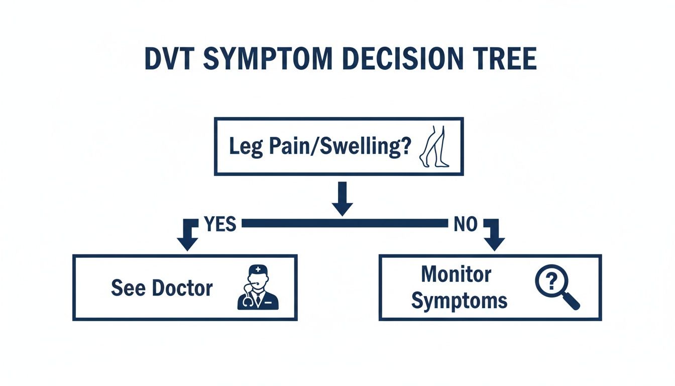
The message is clear: if you have a leg that’s persistently swollen or painful, the safest thing to do is get it checked out by a professional.
The Role of the D-Dimer Test
If your Wells score suggests a low or moderate risk, your GP will likely order a D-dimer test. This is a straightforward blood test that’s brilliant for ruling clots out.
Here’s how it works. When a blood clot forms anywhere in your body, your system naturally starts to break it down. This process releases tiny protein fragments into your blood, and these are called D-dimer. The test simply measures the level of these fragments.
Its real power is in its ability to give you the all-clear.
A negative D-dimer result is incredibly reassuring. It tells us that the chances of you having a significant blood clot are extremely low. In most cases, this means you won’t need any further scans, which can bring immediate peace of mind.
A positive result, however, is a different story. It’s not a confirmation of DVT, just a sign that something is going on. A high D-dimer level tells your doctor that clot formation and breakdown is happening somewhere in your body, but it doesn’t pinpoint where or why. Many other things can cause a raised D-dimer, including:
- Recent surgery or a significant injury
- Infection or inflammation
- Pregnancy
- Some cancers
- Heart conditions
So, a positive D-dimer doesn’t automatically mean you have a DVT. It just means we need to investigate further. It’s the signal that we need to move to the next step, which is usually an ultrasound scan. At The Lagom Clinic, we help arrange rapid access to essential diagnostics like these. You can learn more on our private blood test page.
Visualising the Veins with a Duplex Ultrasound
If your doctor’s initial assessment and your D-dimer test results suggest a DVT could be the culprit, the next move is to get a direct look inside your veins. This is where the compression duplex ultrasound comes in. It’s the gold standard for getting a clear, definitive picture, and it’s completely non-invasive.
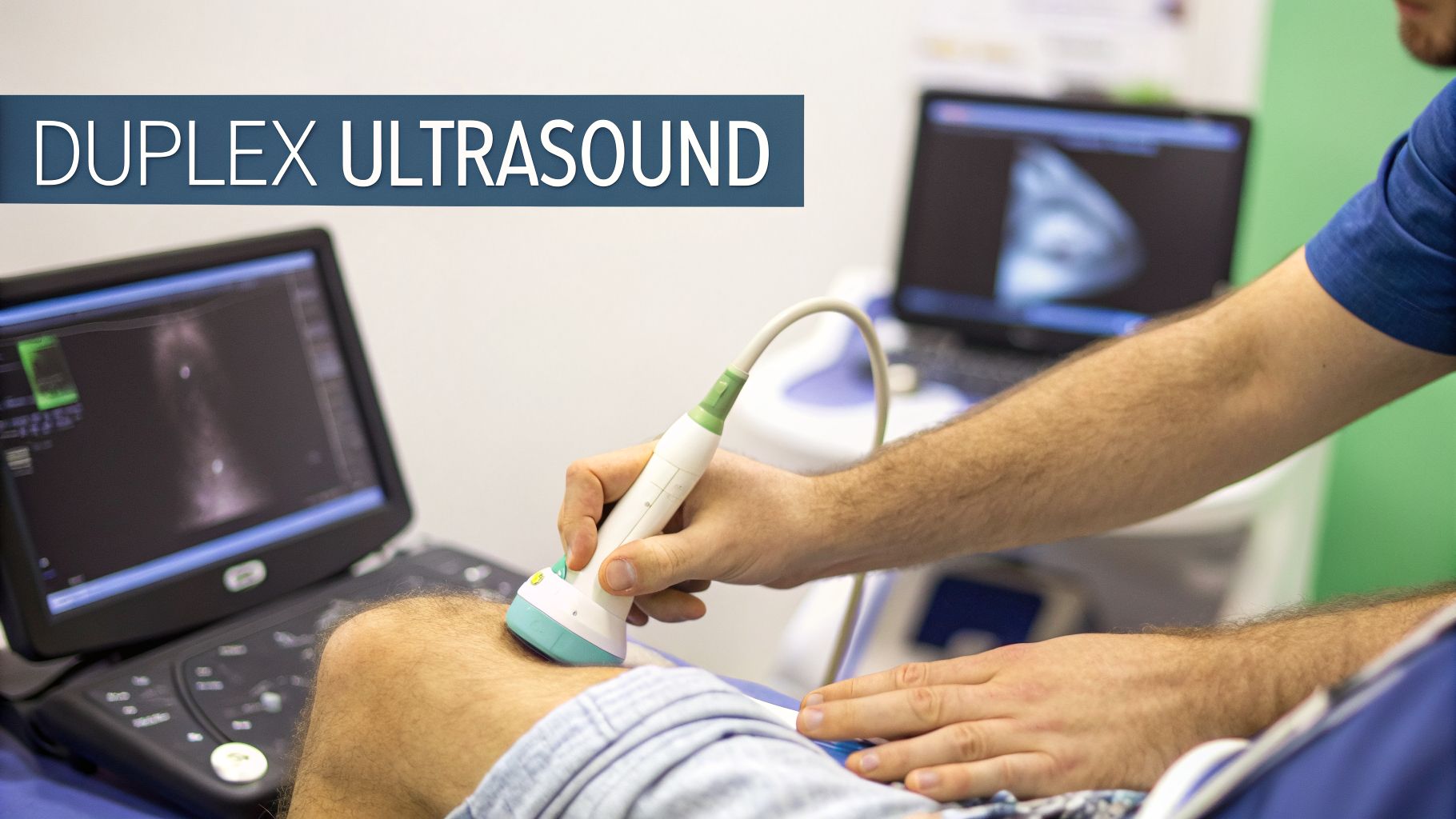
This test is entirely safe and painless, relying on sound waves, not radiation, to see what’s going on. You can think of it as a two-in-one investigation, where two different types of ultrasound work together to give us the full story.
Seeing and Hearing Your Blood Flow
The “duplex” in the name simply means the machine is doing two jobs at once. First, a standard ultrasound generates a black-and-white image, allowing the sonographer—the specialist performing the scan—to visualise the physical structure of your veins.
Simultaneously, the machine uses Doppler ultrasound. This is the “hearing” part of the test. The Doppler technique is brilliant because it can detect movement, showing the sonographer not just if blood is flowing, but in which direction and how quickly. This flow is often displayed in colour on the screen (usually red and blue), making any blockages or disruptions immediately obvious.
What to Expect During the Scan
The whole process is very straightforward and typically lasts between 30 and 60 minutes. You’ll lie down comfortably while a clear, water-based gel is smoothed onto your leg. This gel isn’t medication; it just ensures the ultrasound probe makes solid contact with your skin for the clearest possible image.
The sonographer then gently places a small, handheld probe (a transducer) on your skin, moving it along the major veins in your leg. You’ll feel some light pressure as they do this, which is actually a key part of the diagnostic process.
The most crucial part of the scan is a technique called compression. The sonographer will press down firmly but gently with the probe at different points along the vein. A healthy, clear vein is like a soft, flexible tube and will easily compress flat. A vein blocked by a firm clot, however, will not.
This simple physical check gives an immediate and clear indication. If a vein doesn’t squash down, it’s a very strong sign that a clot is inside. The Doppler’s ‘hearing’ then adds another layer of confirmation by showing if blood flow is being blocked or forced to find a new path around the obstruction.
The accuracy of this deep vein thrombosis test is excellent. For clots in the large thigh veins (above the knee), it’s more than 95% sensitive.
What the Ultrasound Can Tell Your Doctor
A duplex ultrasound provides far more than a simple “yes” or “no”. It gathers a wealth of detailed information that is vital for planning your treatment.
- Confirmation: It can definitively confirm the presence of a DVT.
- Location: The scan pinpoints exactly which vein the clot is in.
- Size: It helps the clinical team understand the size of the clot and how much of the vein is obstructed.
- Blood Flow: It reveals how the clot is affecting circulation throughout your leg.
This level of detail empowers your doctor to make the best decisions for your care. It not only confirms the diagnosis but also helps gauge the severity and provides a crucial baseline for monitoring how well treatment is working. At The Lagom Clinic, we arrange rapid access to these essential scans, ensuring our patients get a fast diagnosis and can start the right treatment without unnecessary delays.
When We Need a Deeper Look: Advanced DVT Imaging
A duplex ultrasound is fantastic for finding clots in the more accessible veins of the leg, but it has its limits. Sometimes, we suspect a clot is lurking deeper in the body—in the large, central veins of the pelvis or abdomen, for instance. In these tricky situations, or if an ultrasound gives us an inconclusive result, we need to bring out the big guns.
This is where advanced imaging comes in. These techniques give us a highly detailed, three-dimensional ‘roadmap’ of your veins, leaving no stone unturned. They’re not the first port of call, but they are essential when the initial picture isn’t clear enough.
Creating a 3D Roadmap of Your Veins
The two main advanced scans we rely on are CT venography and MR venography. They work differently—a CT scanner uses specialised X-rays, while an MRI machine uses powerful magnets—but they share the same goal: to create a crystal-clear, cross-sectional image of your veins.
To get this incredible detail, both scans typically involve a special contrast dye. This is injected into a vein (usually in your arm) and travels through your bloodstream. As it circulates, the dye makes your veins ‘light up’ on the scan, vividly outlining their structure and showing any blockages with remarkable precision. The time an MRI takes can vary, so it helps to be prepared. We cover this in more detail in our guide on how long an MRI takes.
Think of it like pouring a brightly coloured liquid into a complex network of clear pipes. Suddenly, you can see the exact path the liquid follows and pinpoint exactly where it’s being blocked. That’s what these scans do for your veins.
For you, the experience is quite simple. A CT scan involves lying on a bed that slides through a large, ring-shaped scanner. An MRI machine is more of a short tunnel. For both, the most important thing is to stay as still as you can, which helps us get the sharpest possible images.
The Original Gold Standard: Contrast Venography
Before CT and MR scans were so advanced and widely available, the definitive test was a contrast venography. This is a more hands-on procedure where dye is injected directly into a vein in your foot. A series of X-rays then tracks the dye’s progress as it travels up the leg.
It’s an incredibly accurate test. However, it’s also more invasive than a CT or MRI and exposes you to a bit more radiation. Because of this, we don’t use it nearly as often these days.
That said, it still has a role to play. In particularly complex cases, or when other imaging methods aren’t an option, contrast venography remains a reliable backup. It ensures that even in the most challenging situations, we have a way to get the clear answers needed to protect your health.
Taking Control: Proactive Lifestyle Changes to Lower Your DVT Risk
Once you’ve been through the process of a deep vein thrombosis test, your focus naturally shifts to the future. Knowing you can actively reduce your risk of another clot is an incredibly empowering part of taking control of your health. The good news is that simple, consistent lifestyle adjustments can make a world of difference to your vascular wellbeing.
These proactive habits are your best defence. They work by keeping your blood flowing smoothly and your veins in the best possible condition. Best of all, they are practical, accessible, and based on solid medical evidence.

Embrace Consistent Movement
If there’s one golden rule for preventing DVT, it’s this: keep moving. Long periods of sitting or standing still are a clot’s best friend. When you’re immobile, blood can pool in the deep veins of your legs, creating the perfect environment for a DVT to form. The solution is beautifully simple—move more.
Regular physical activity is fantastic for your circulation. This doesn’t mean you need to start training for a marathon. A brisk 30-minute walk on most days of the week is hugely beneficial. If you have a desk job or you’re on a long flight, make a conscious effort to get up and walk around for a few minutes every single hour.
Even tiny movements from your chair can help:
- Ankle Circles: Lift your feet clear of the floor and gently rotate your ankles, first clockwise, then anti-clockwise.
- Calf Raises: With your feet flat, lift your heels up high, then lower them. Reverse the movement by keeping your heels down and lifting your toes.
- Foot Pumps: Imagine you’re pressing a pedal. Point your toes down towards the floor, then pull them up towards the ceiling, alternating between feet.
These small exercises might not feel like much, but they contract your calf muscles, which act as a ‘second heart’ to pump blood up and out of your legs, preventing it from stagnating.
Maintain a Healthy Body Weight
Carrying extra weight puts constant pressure on the veins in your legs and pelvis. Think of it like a roadblock for your circulation. This pressure can damage the delicate one-way valves inside your veins that are designed to keep blood flowing towards the heart. When these valves weaken, blood can flow backwards and pool, which seriously increases your DVT risk.
Achieving and maintaining a healthy weight through a balanced diet and regular exercise takes that strain off your veins. It’s one of the most effective long-term strategies you can adopt to protect your entire circulatory system.
Prioritise Proper Hydration
The consistency of your blood matters. When you become dehydrated, your blood naturally gets thicker and more concentrated, which makes it more likely to form clots. Staying well-hydrated is such a simple but critical preventative step.
Make sure you’re drinking plenty of water throughout the day. A good benchmark is to drink enough so that your urine is a pale straw colour. This simple habit helps ensure your blood flows freely and easily through your veins. As you work on these lifestyle changes, you might find these Top Tips On Hydration Nutrition Exercise Sleep a helpful resource.
The Critical Decision to Stop Smoking
If you’re a smoker, quitting is the single most powerful thing you can do for your vascular health. The thousands of chemicals in cigarette smoke actively damage the smooth, delicate inner lining of your blood vessels.
This damage creates a rough, ‘sticky’ surface inside the veins, making it far easier for platelets and other clotting agents to clump together and form a dangerous thrombus. When you quit smoking, your blood vessels can begin to heal, dramatically lowering your risk of DVT and a whole host of other cardiovascular problems.
The Role of Compression Stockings
For some people, compression stockings are an essential tool. This is especially true for those with known risk factors, or during high-risk times like after surgery or on a long-haul flight. Your doctor may recommend wearing graduated compression stockings.
It’s important to understand these aren’t just your average tight socks. They are specifically engineered medical garments that apply the greatest pressure at the ankle, with that pressure gradually easing as it goes up the leg. This gradient squeezes the leg veins, helping to push blood upwards and stop it from pooling.
Your doctor is the best person to advise if compression stockings are right for you. They can also make sure you get the correct size and pressure level for your specific needs, making them a valuable part of your DVT prevention plan.
Building these habits into your daily routine can feel overwhelming at first. To help, here’s a simple breakdown of how to turn these recommendations into action.
Your DVT Prevention Plan Practical Lifestyle Adjustments
| Risk Factor | Preventative Action | Why It Helps |
|---|---|---|
| Immobility | Move for a few minutes every hour. Take the stairs instead of the lift. Do simple leg exercises while seated. | Contracts calf muscles, pumping blood out of the legs and preventing pooling. |
| Excess Weight | Aim for a balanced diet and regular exercise, like a 30-minute daily walk. | Reduces the constant physical pressure on your leg veins, protecting their internal valves. |
| Dehydration | Drink plenty of water throughout the day. Aim for pale, straw-coloured urine. | Keeps blood at a healthy consistency, making it less 'sticky' and prone to clotting. |
| Smoking | Seek support to quit. Talk to your GP about cessation aids and programmes. | Allows the damaged lining of your blood vessels to heal, making them less prone to clot formation. |
| High-Risk Situations | Wear properly fitted graduated compression stockings during long travel or post-surgery. | Provides external pressure to assist veins in pushing blood back towards the heart. |
Making even one or two of these changes is a positive step forward. The key is consistency—turning small, positive actions into lifelong habits for better vein health.
Common Questions About DVT Testing
Going through the process of being tested for a deep vein thrombosis (DVT) can naturally bring up a lot of questions. Knowing what to expect, how long it all takes, and what happens next can make the entire experience feel much less stressful.
We’ve put together some clear, straightforward answers to the questions we hear most often, helping you feel more informed and in control.
How Long Will I Wait for My DVT Test Results?
The time it takes to get results back really depends on which specific deep vein thrombosis test you’ve had. The good news is that the first-line tests are very quick.
- D-dimer Blood Test: This is the fastest of the lot. Results are often back from the lab within a few hours, giving your doctor a quick indication of whether further investigation is needed.
- Duplex Ultrasound: During the scan itself, the sonographer can often see in real-time if there's a clot. A formal, detailed report is then usually prepared and sent to your doctor the very same day.
- CT or MR Venography: Because these are much more detailed scans, they need a specialist radiologist to carefully analyse all the images. You can typically expect the full report to reach your doctor within 24 to 48 hours.
The whole diagnostic process is designed to be efficient. The priority is always to get a definitive answer as quickly as possible so you can either get the all-clear or start treatment without any delay.
Are the Diagnostic Tests for DVT Safe?
Absolutely. Patient safety is always the number one priority, and the main tests used to look for a DVT are extremely safe.
A D-dimer test is just a standard blood draw, with only a tiny risk of a little bruising where the needle went in. The duplex ultrasound, which is the go-to imaging tool, is completely non-invasive. It uses harmless sound waves—no radiation at all—making it perfectly safe for everyone, including expectant mothers.
The more advanced scans, like a CT scan, do involve a low dose of radiation, but the amount is carefully managed to be as minimal as possible. Both CT and MR venography might also use a special contrast dye to make your veins show up more clearly. There's a very small risk of an allergic reaction to this dye, but your medical team will always go through your health history (including how well your kidneys are working) and discuss any potential risks with you before going ahead.
What Happens If a DVT Is Found on My Test?
Hearing that you have a DVT can be worrying, but it’s so important to remember that it's a very treatable condition with modern medicine. If a test does confirm a clot, your doctor will get you started on treatment straight away.
The main treatment for a DVT is anticoagulant therapy, which you might know as blood thinners. These medicines are crucial because they stop the clot from getting any bigger and prevent new ones from forming. This gives your body the chance to gradually break down and absorb the clot over time.
Treatment usually lasts for at least three months, but the exact duration will depend on your specific situation. Your doctor will talk you through the different medication options—which might be daily tablets or injections—and will keep a close eye on your progress.
Can a DVT Diagnosis Affect My Travel Plans?
This is a very practical and common question. After going through a deep vein thrombosis test and getting a diagnosis, many people wonder how it might impact their travel plans. A key part of this is understanding your options for travel insurance with pre-existing conditions. It’s always best to have a chat with your doctor before you book any long-haul flights or trips.
Your doctor can give you personalised advice on when it’s safe for you to travel and what simple precautions you should take to stay healthy on the move.
How Can I Reduce My DVT Risk During Long Journeys?
Being proactive is the best way to protect yourself during long periods of sitting still, whether you're on a plane, a train, or in a car.
- Move Regularly: Try to get up and have a little walk around the cabin or carriage every hour or two. If you're driving, plan for regular stops to get out and stretch your legs.
- Do Seated Exercises: If you can't get up, simple leg exercises from your seat make a real difference. Flex your ankles, point your toes, and do some calf raises to keep the blood flowing.
- Stay Hydrated: Drink plenty of water. It's best to avoid too much alcohol or caffeine, as they can cause dehydration, which can make your blood a bit thicker.
- Wear Compression Stockings: Your doctor might suggest wearing properly fitted compression stockings, which give your circulation a helpful boost during travel.
These simple steps put you in control, helping you manage your risk so you can travel with more confidence and peace of mind.
At The Lagom Clinic, we understand that when you have a health concern, you need clear, compassionate answers without the wait. Our private GP service in Bristol is set up to provide rapid assessments and fast-track access to diagnostics, including blood tests and imaging. To book a consultation and take the first step towards clarity, visit us at https://www.thelagom.co.uk.


 Members Consultation